


A group of young scientists, including staff, graduate students and students of the Department of Electronics at the National Research Nuclear University MEPhI, is creating a PixelVision pixel detector capable of detecting individual X-ray photons and measuring their energy. This will allow obtaining data for a detailed analysis of the composition of materials and biological tissues, as well as forming "color" X-ray images. The project team is currently participating in the fourth cycle of the Rosatom and MEPhI University Technology Accelerator.
Traditional radiography is based on varying degrees of absorption of X—rays by body tissues: dense structures (for example, bones) delay radiation more strongly, soft tissues - weaker. This creates a contrasting black and white image where the brightness corresponds to the absorption level. However, this method does not allow analyzing the energy of individual photons. The development of technologies that record the spectral characteristics of radiation is a key step towards creating a "color X—ray" capable of improving diagnostics. There are no such solutions in Russia yet, and the Pixel Vision project, implemented with the support of experts from TVEL JSC (Rosatom), is designed to fill this gap. The scientific consultant is Eduard Atkin, head of the Laboratory for the Design of specialized integrated circuits at the Department of Electronics at the National Research Nuclear University MEPhI.
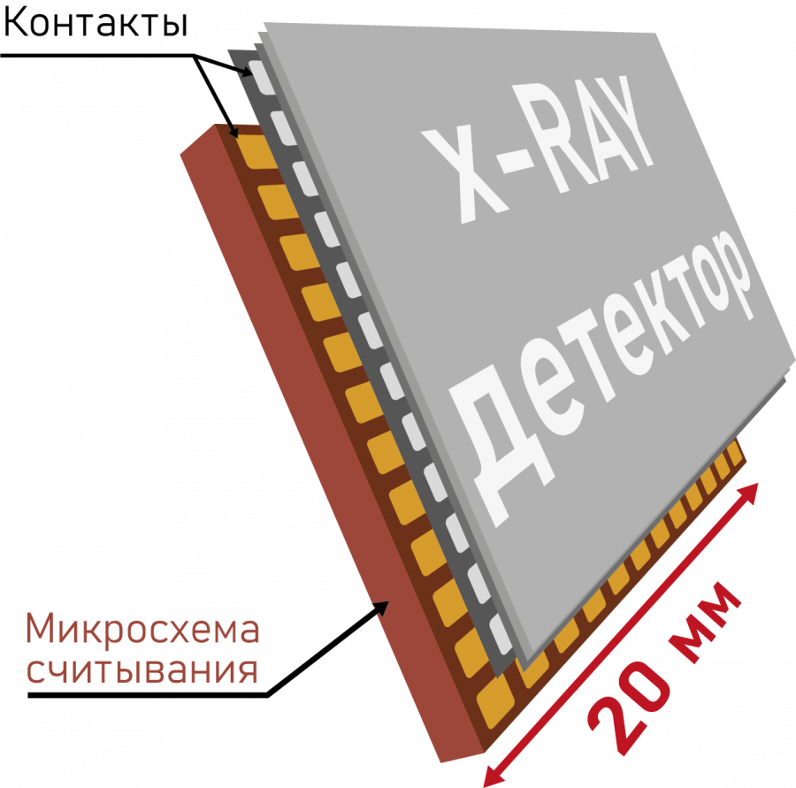
As explained by the head of the PixelVision project, graduate student at MEPhI Danila Lobankov, the detector is a two-layer structure. The top layer is a gallium arsenide sensor developed at Tomsk State University. When X-ray photons interact with the sensor material, ionization occurs, leading to the formation of electron-hole pairs. Their number is proportional to the energy of the absorbed photon.
The bottom layer is a specialized microcircuit created at MEPhI. Its task is to read, process charges and convert signals into data on the spatial distribution and energy of photons. It is planned that the 20×20 mm chip will contain 65,536 pixels (256×256). At the first stage, the developers will create a prototype with a 32x32 pixel matrix.
The key difficulty is the high sensitivity of the device. The magnitude of the charges detected by the sensor is on the order of several thousand electrons (~1.6 ×10⁻1⁶ Kl). To work with such signals, the microcircuit is designed using CMOS technology with standards of 180 nm.
As noted by the project participant, MEPhI graduate student Salavat Yamaliev, the introduction of the detector into medicine will not only allow obtaining high-definition "color" X-ray images, but also reduce the radiation dose to patients by reducing exposure time. The technology will also find applications in scientific experiments (megascience class installations), inspection systems and industrial non-destructive testing.
By the beginning of 2026, the PixelVision team plans to complete the development of a prototype detector and prepare a business model for the project. The project participants are currently undergoing a training program at the University Technology Accelerator with the participation of experts and potential customers from Rosatom. And next summer, together with other participants of the program, the authors of the development will compete for investments of up to 5 million rubles for the development of their business.