


Researchers of the Laboratory of Nano-Bioengineering at PhysBio in collaboration with colleagues have proposed an original approach to nanoscale 3D-analysis of materials, successfully testing it on an innovative own-produced installation.
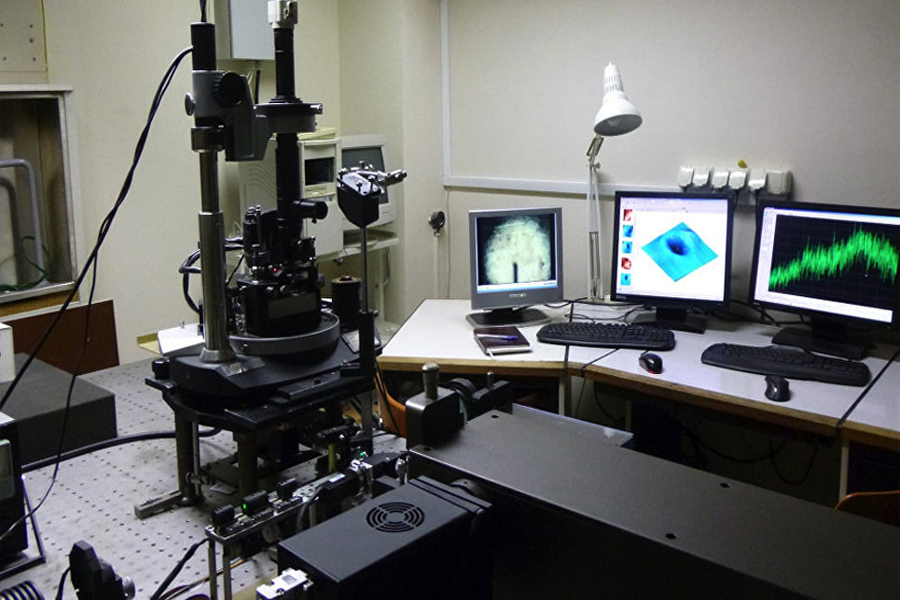
General view of the device in the laboratory
The authors managed to combine all the advantages of various modern approaches to nanoscale measurements in one unit: scanning probe microscopy (analysis of surface and physical parameters of the object), optical microspectroscopy (chemical mapping and determining the optical properties) and nanotomography (accurate 3D visualization of the internal structure of the object based on the plurality of x-ray images). This combination of methods allows, in addition to high-quality 3D images of material’s nanoscale parts, to register the spatial distribution of its mechanical, electrical, optical and chemical properties (e.g. elasticity, conductivity, magnetization) in these same areas.
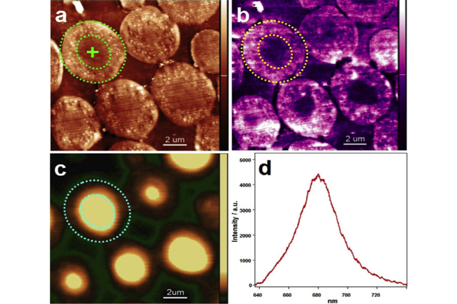
An example of data of a nanoscale analysis, obtained by the combination of 3D scanning probe nanotomography and optical microscopy
The creators have successfully tested their project in a comprehensive study of the fluorescent-labeled polymer microspheres used in modern immunodiagnostics, both for multiparameter detection of markers of various diseases and personalized medicine for the detection of rare events, such as the appearance of circulating cancer cells and micrometastases.
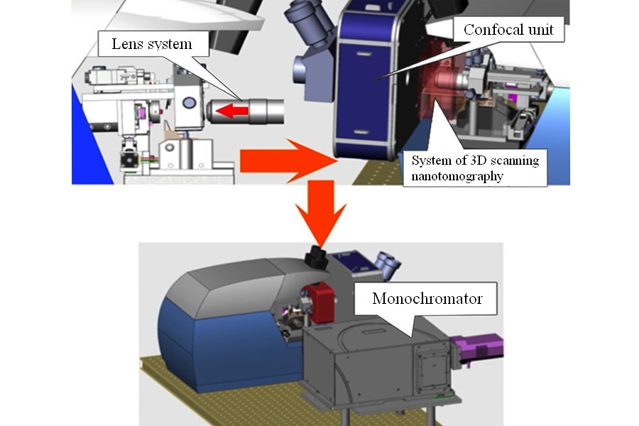
System for integrated 3D analysis of nanomaterials. At the top left – a sketch of the combined system of the 3D scan nanotomography and optical microscopy. At the top right – diagram of a deivce, which includes the confocal unit, lens system and the 3D-scanning nanotomography. Below – general schematic view of the combined system (in section).
The leading scientist of the Laboratory of Nano-Bioengineering (LNBE) MEPhI, doctor of chemical sciences, Professor Igor Nabiev noted: "This instrumental approach retains all the advantages of scanning microscopy and optical microspectroscopy, allowing to obtain multi-parametric 3D feature with the effective combination of both methods. The results of the study can be used for successful conversion of analysis data from 2D to 3D, obtained using most methods of optical probe nanoscopy implemented with modern high-resolution devices.”
This development can be used for comprehensive analysis of biological samples. In addition, it opens new possibilities in the field of quality control in the creation of defect-free nanomaterials, systems for targeted delivery of drugs using nano-sized "containers" and also in solution of nanosafety problems and associated challenges of determining nanoparticles penetration in different organs and tissues of a living organism.
Photo: © Fotolia / Photokanok_1984